Limb-to-crotch. Repost

Israel Sarrió arrived at the Rehabilitation Center of Levante from Hospital Peset in Valencia (Spain) on January 31st of 2004 with an amputation of the left arm 2 inches above the elbow.
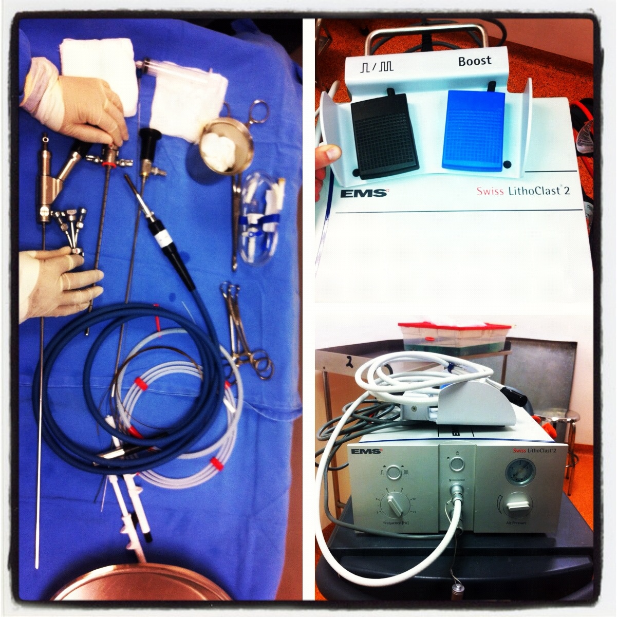
Amputated ArmTrunnionrx
Click on images for large size
At 14:00hrs, the microsurgery implant began. The first maneuver consisted in lavage and sterilization of the amputated arm and its connection to the brachial artery by a silicone tube in order to revascularize it.

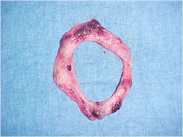
Later we rebuilt the humerus and repaired the brachial artery, the basilic and cephalic veins, and the three nerves of the arm.

This proccess lasted 21 hours, and the reimplantation was successful. The patient went to the Intensive Care Unit during that night and the next day, he went to a normal room.

first implant
Two days later, we found a wound infection that was seriously compromising the viability of the reimplantation. We talked to the family and they told us that the day of the accident the arm fell in a drain. This contamination was the probable cause of the unfavorable progress. The situation was critical, and we said to the family that there were two different options: reamputate the arm or try to save it by taking it into a healthy zone where it would be possible to nourish it while we cleaned the infected area.

wound infection
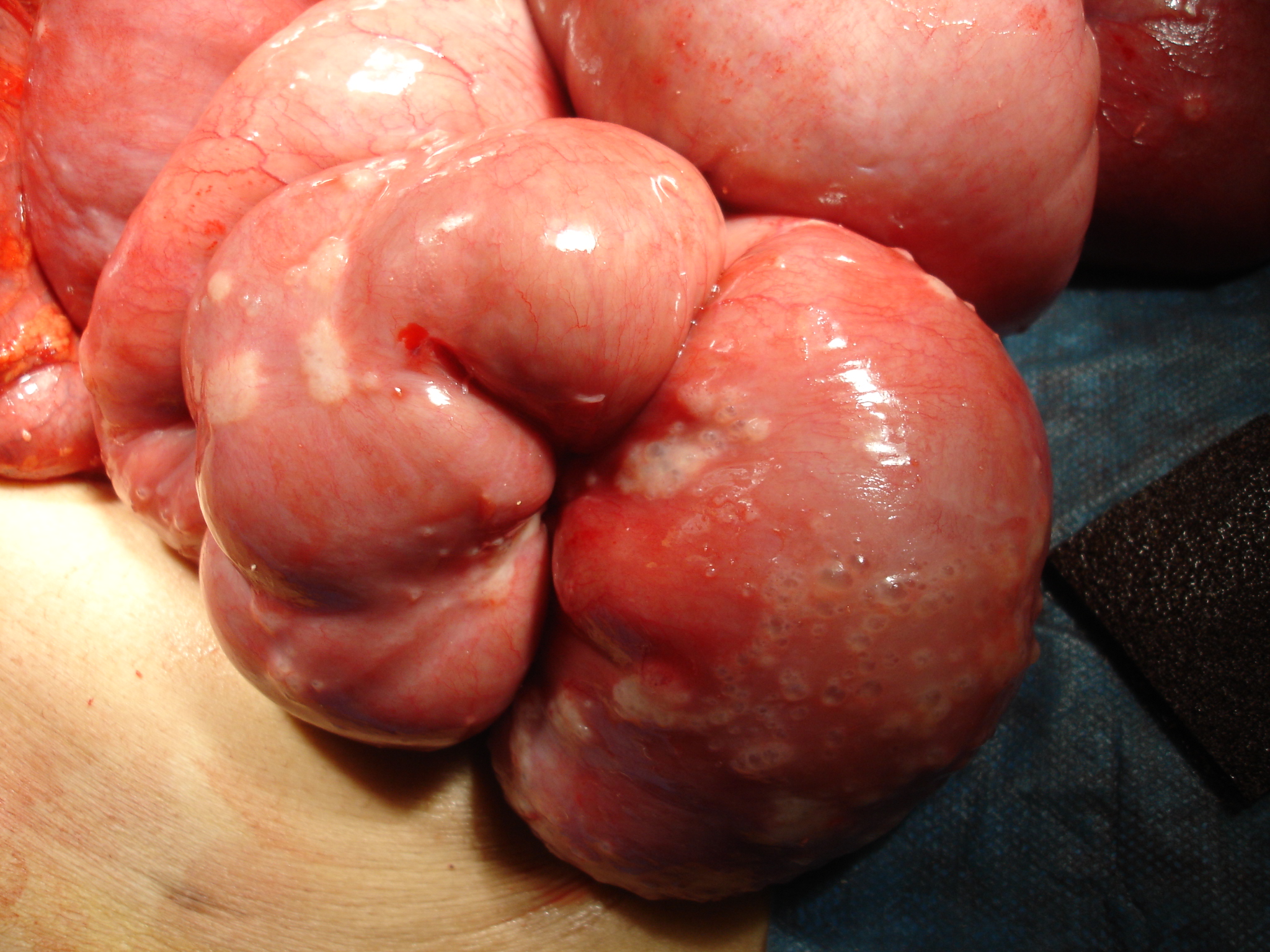
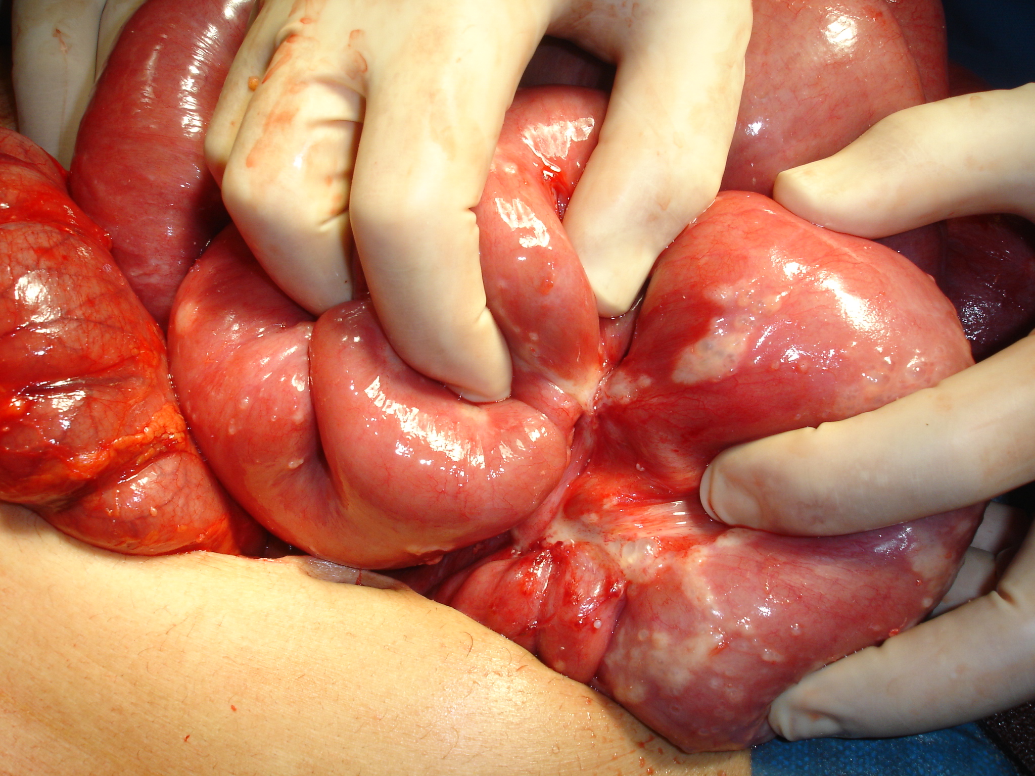
The idea of taking the arm to another anatomical location came by reading a similar case published by Michael Wood (Mayo Clinic, Rochester, MN, USA) in which he attached an arm in the groin. The uniqueness of our case consisted of using the procedure to rescue the limb from an infection, called a Deferred Transitory Heterotopic Implant. This was the first such case of involving an elbow.

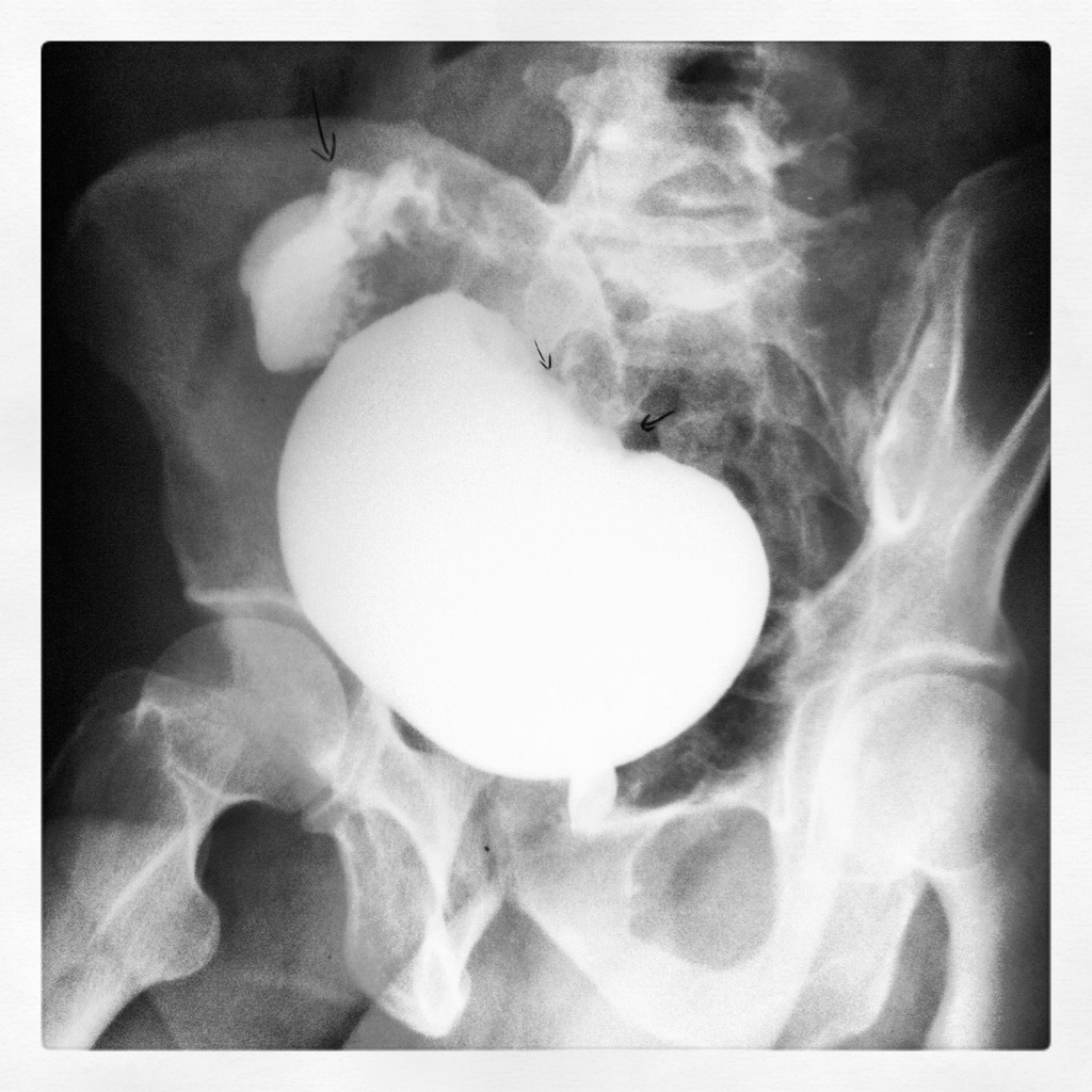
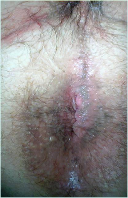
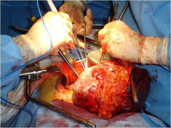
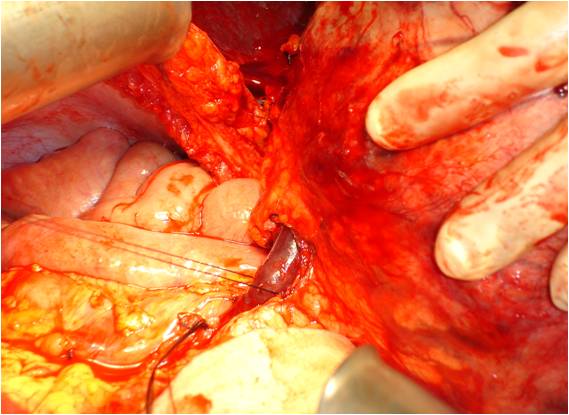
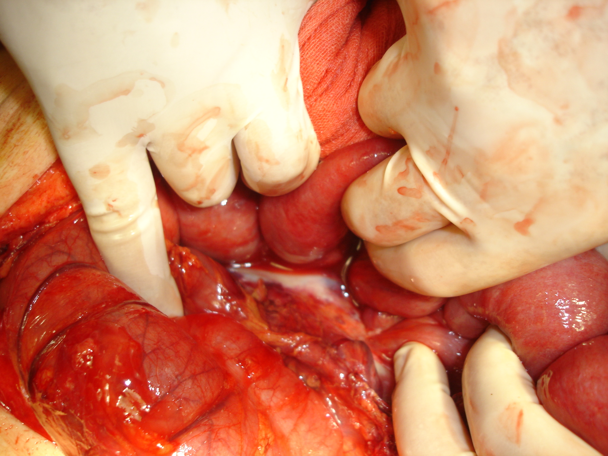
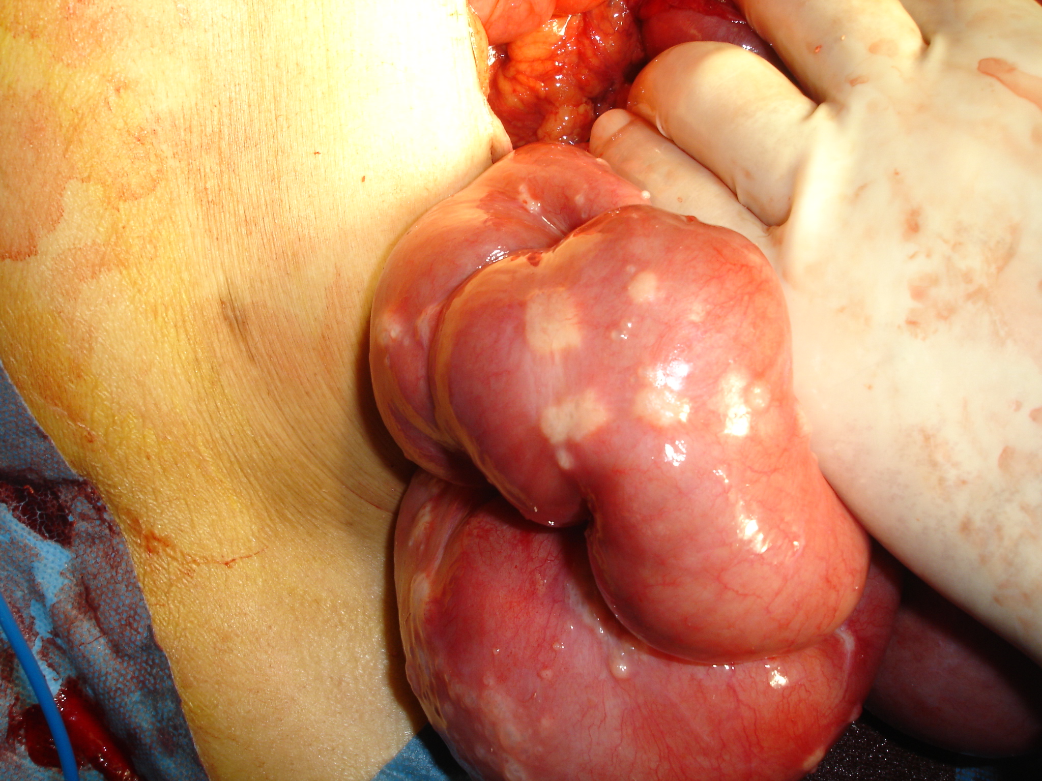
With the agreement of the family it was decided to transfer the arm to the groin where large blood vessels are readily accessible. In 4 hours we disassembled the reconstruction performed earlier and connected the blood vessels of the arm to those of groin by means of microsurgery.

implant in dangerleg implantleg implantleg implant
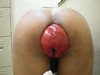
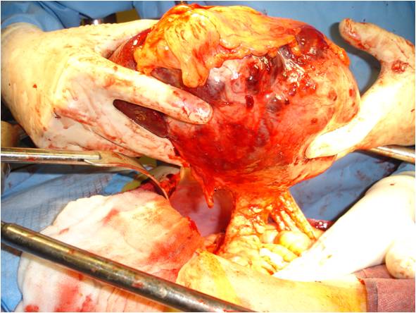
The psychological impact on the patient was very important. As soon as he woke up, we told him that the arm was infected and we have to save it in other location on his body. After a couple of hours he realized that the arm was on his leg. He stayed that way for nine days with daily cleanings until it was verified that the stump was clean.

On February 12nd of 2004, we reimplanted the arm in its original place, rebuilding bone, arteries, veins, nerves, muscles and skin in a procedure of 6 hours.

replantpost replant
Afterwards, the patient was doing physically and psychologically well, happy that he have not have to lose his arm.

By this time the patient could move his elbow. From the time of the surgery, the repaired nerve functionality is growing at a speed of 1 milimeter per day. In about 6 months we hope that he will be able to move his hand and fingers.

The function which we hoped to obtain from this procedure was better than those obtained with an amputation and the prosthesis. The patient will be able to grasp objects, to perform normal life functions, such as tie his shoes, all with small orthopedic aids.

final status
The complete surgical team of the Unit of Hand and Reconstructive Surgery was involved in this operation, with Dr. Pedro Cavadas leading the team, and Dr. Navarro, Dr. Soler, Dr. Duke and Dr. Landin as assistants.

Via

Recto-Vesical Fistula

20120229-130702.jpg

Recto-vesical fistula is an anomalous communication (fistulous tract) between the bladder and the rectum. The common etiologies are diverticulitis, Crohn’s disease, irradiation, traumatic, bladder or colo-rectal cancer and tuberculosis.

Common symptoms of presentation are fecaluria (presence of feces in the urine), pneumaturia (passage of gas in the urine), hematuria (presence of blood or clots in the urine), intestinal symptoms, abdominal pain, and fever/chills.

Evaluation: urine test (urinalysis) shows food fibers, fecal particles, bacteria, white blood cells and red blood cells.

Cystography shows fistulous tract and bladder inflammation, as seen in this picture, which is a diagnostic/evaluation method barely used because of availability of CT scans.

Later I will give you the fast facts about treatment of this condition.

Rectal Prolapse – NSFW

Rectal prolapse Rectal prolapse Rectal prolapse

As in Marvin L. Corman’s book (Colon & Rectal Surgery) quotes:

Man should always strive to have his intestines relaxed all the days of his life and that bowel function should approximate diarrhea. This is a fundamental principle in medicine, that whenever the stool is withheld or is extruded with difficulty, grave illnesses result.

Maimonides: Mishneh Torah

Rectal prolapse (a.k.a. Procidentia) is one of the most fascinating surgical pathologies because its complexity in treatment. In 1912 Moschcowitz proposed an herniation of Douglas pouch as a cause for rectal prolapse. Another cause was proposed by Broden and Snellman with the help of defecography described a full thickness rectal intussusception.

Rectal prolapse is 6 times more common in females (as males) aged 50 years or older. Many of male patients has a past medical history of psychiatric disorders. Chronic or lifelong constipation with straining is present in more than 50% of patients.

Patients describe a mass or bulge that they have to push back in after defecation. Often, presentation of rectal prolapse can be dramatic when the prolapsed segment becomes incarcerated below the level of the anal sphincter as in this clinical case and emergency surgical therapy was indicated.

More than fifty types of procedures for repair rectal prolapse have been described, but the treatment is always surgical.

Succesful surgery in a Girl with eight limbs

Lakshmi is the hindu goddess of of wealth, fortune, love and beauty, the lotus flower and fertility. She is represented by an icon of a lady with four arms.

Lakshmi Tatma is a girl who is two years old but she was born with 8 limbs, because she is joined at the pelvis to what is, in fact, a headless, undeveloped twin (parasitic twin or to be more specific, ischiopagus cojoined twin).

This is surely an abnormal feature and it has attracted media attention worldwide.

The 24-hour-long surgery to remove the extra limbs of this Indian girl born with four arms and four legs was a success, doctors announced Wednesday.

A team of more than 30 physicians removed Lakshmi’s extra limbs, salvaged her organs, and rebuilt her pelvis area, Dr. Sharan Patil said from a hospital in the southern Indian city of Bangalore, India.

Surgical model

This surgical model is made of glass fiber and has abdominal and thoracic compartments separated. A selective bronchial intubation can be done in this model. It also has a structure that simulates a diaphragm. You can perform both laparoscopy and thoracoscopy.

The organs of a pig are placed within the model.


The model is intubated with an “orotracheal” tube

abdominal view
Abdominal view

thoracic view
Thoracic view

Laparoscopy
Laparoscopy view

Hemorrhoidal prolapse & PPH procedure

This is a case of a 67 years old male with history of chronic constipation (20 years) and chronic hemorrhoidal disease came to the office with rectal bleeding and a non-redicible anal mass.

On physical examination we found Grade IV hemorrhoidal disease.

hemorrhoidal prolapse

We decide to perform a PPH procedure with an hemorrhoidal circular stapler (33 mm) kit.


Note the tissue removed.


This donut confirms the circular and complete resection of the defect.

On the postoperatory period the patient was in excellent condition without pain and without bleeding.

Pancreatic neoplasm

A previously healthy 27 years old female came to the emergency department with a chief complaint of one-month diffuse and intermitent abdominal pain, transitory relief with bowel movements, nausea without vomit, early saciety, postprandial fullness and weight loss of 4 kilos in 1 month.

On the physical exam, we found a 10×15 cm tumor in the left upper quadrant. The tumor is not fixed, it feels solid and well delimited. We did not find any other important data.

Lab work beteween normal ranges. We ordered plain abdominal film, chest x ray and later an abdominal ultrasound and a CT scan.

Rx

Ultrasound

CT Scan

With these findings, we decided to perform an exploratory laparotomy (left subcostal incision) and tumor resection.


Look the size of the tumor and its characteristics.


Note the splenic vein.


The tumor arises from the tail of the pancreas. It was impossible to dissect the spleen, so we had to cut it.

Pathology

Final diagnosis: Benign Solid Pseudopapillary Tumor of the Pancreas of 14 cm, and 1.66Kg.

Solid-pseudopapillary tumor (SPT) of pancreas is a very rare (about 450 cases have been described in the world literature) neoplasm of low malignant potential and unknown origin. It generally occurs in young women and the prognosis is usually good after complete surgical removal.

SPT is also know as Fratz’s tumor

Bochard triad

A 68 year old female patient came to the E.D. with a chief complaint of sudden, severe epigastric pain, retching without vomitus and distention of the upper abdomen.

The patient was alert, very anxious, and in severe pain.
blood pressure of 160/120; pulse of 126; respiratory rate of 24; normal temperature; SatO2 of 96% without O2 supplement.
She had a distended upper abdomen and epigastric tenderness without rebound or guarding.

A plain film of the abdomen was ordered and showed gas-filled, grossly dilated stomach.

Pass of nasogastric tube was unsuccessful.

A laparotomy was mandatory.

The patient survived the surgery but 6 months later, she died by causes nonrelated to the surgery

In 1904, Borchardt described the classic triad of gastric volvulus: severe epigastric pain, retching without vomiting, and inability to pass a nasogastric tube.